
Dog Paw Pads play a crucial role in the locomotion of digitigrade animals like dogs and cats, absorbing shocks from the ground to protect joints and muscles. Unlike human heels, the dog paw pad structure features a sophisticated multi-layered design that excels at cushioning impacts during running or jumping. This article dives into the micro-scale characteristics revealed by recent scientific research on German Shepherd and Chinese Rural dogs, explaining how these layers work together for superior shock absorption. Understanding this can help pet owners better care for their dogs’ paws, preventing injuries from rough terrains.
Digitigrade mammals, such as dogs, walk on their toes with elongated bones and specialized pads under the digits and metacarpals. These pads endure ground reaction forces (GRFs) up to six times a dog’s body weight, especially in active breeds like German Shepherds. Without effective cushioning, these forces could lead to joint diseases or musculoskeletal injuries. Past studies focused more on human or plantigrade feet, but this research uses scanning electron microscopy (SEM), histology, and finite element (FE) analysis to uncover the dog paw pads cushioning secrets at a microscopic level.
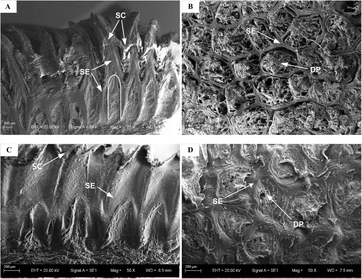 Scanning electron microscope images showing spike-like structures and honeycomb pits in the stratified epithelium of dog paw pads from German Shepherd and Chinese Rural dogs
The Multi-Layered Anatomy of Dog Paw Pads
The dog paw pad consists of three main layers: the epidermis (outermost), dermis (middle), and subcutaneous (innermost). The epidermis includes a stratified epithelium layer topped with a tough stratum corneum and embedded dermal papillae, forming a unique honeycomb structure. This is only present on the bottom surface contacting the ground, not side walls.
Histological exams reveal spike-like projections on the outer surface for grip and traction, with internal pits filled by dermal papillae—soft protrusions from the dermis. The dermis features collagen fiber bundles and elastic fibers, acting as a bridge. The subcutaneous layer is packed with adipose tissue divided into small compartments by collagen membranes, resembling a hydrostatic system that stores and dissipates energy.
This setup ensures the paw pad withstands wear, friction, and high-impact GRFs. For pet owners, recognizing these layers highlights why cracked or worn pads need prompt care—regular moisturizing and avoiding hot pavements preserves the natural cushioning.
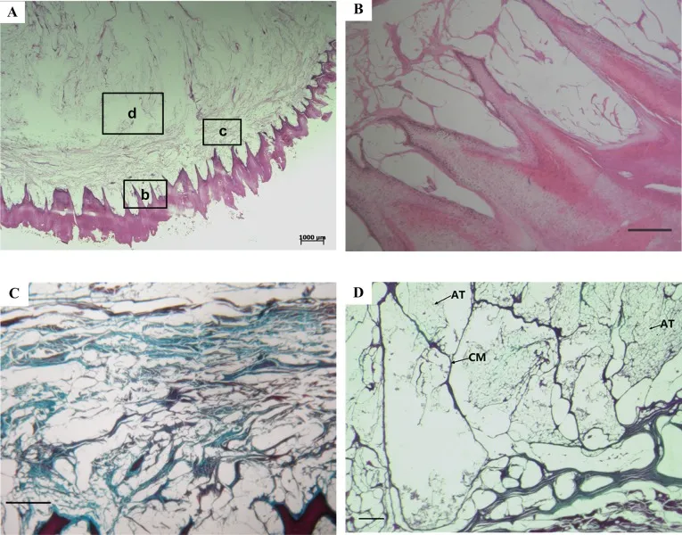 Histological cross-sections of dog paw pad layers, showing stratified epithelium, dermal papillae, collagen in dermis, and compartmentalized adipose tissue in subcutaneous layer
The honeycomb epidermis effectively distributes stress. SEM images confirm a 1:1 ratio of spikes to pits, with holes arranged honeycombed for optimal energy dissipation.
To support your dog’s active lifestyle and protect those vital paw pads during rest, consider a comfortable dog beds near me option that mimics soft ground cushioning.
Finite Element Analysis: Proving the Cushioning Power
Researchers built micro-scale FE models of the epidermis layer—a cube with honeycomb holes filled by dermal papillae cylinders—simulating impacts from 0.05 to 0.4 m/s, typical of dog walking speeds. A structured model (honeycomb) was compared to a uniform one (solid epithelium).
At 0.4 m/s, the structured model reduced peak GRF by 37% (1.7 N vs. 2.7 N) and increased contact time (0.0009 s vs. 0.0006 s), allowing gradual force absorption. Vertical displacement rose 42%, but remained minimal (max 0.23 mm), not hindering speed.
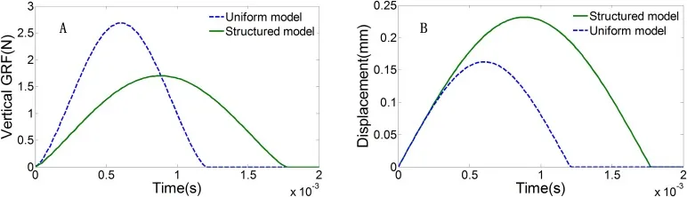 Time histories of vertical GRF and top plate displacements in structured vs. uniform models of dog paw pad epidermis during impact
Von Mises stress analysis showed dramatic drops along dermal papillae (from 1.218 MPa at bottom to 0.00032 MPa at top in structured model), versus slower decay in uniform. Stress concentrated less in the structured version, uniformly distributing in epithelium to prevent hotspots.
Across velocities, peak GRF rose linearly but less steeply in structured models (ratios dropping to 0.63 at high speeds). Peak stresses were consistently lower, confirming superior cushioning.
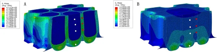 Von Mises stress distributions under peak GRF at 0.4 m/s impact velocity in structured (A) and uniform (B) dog paw pad models
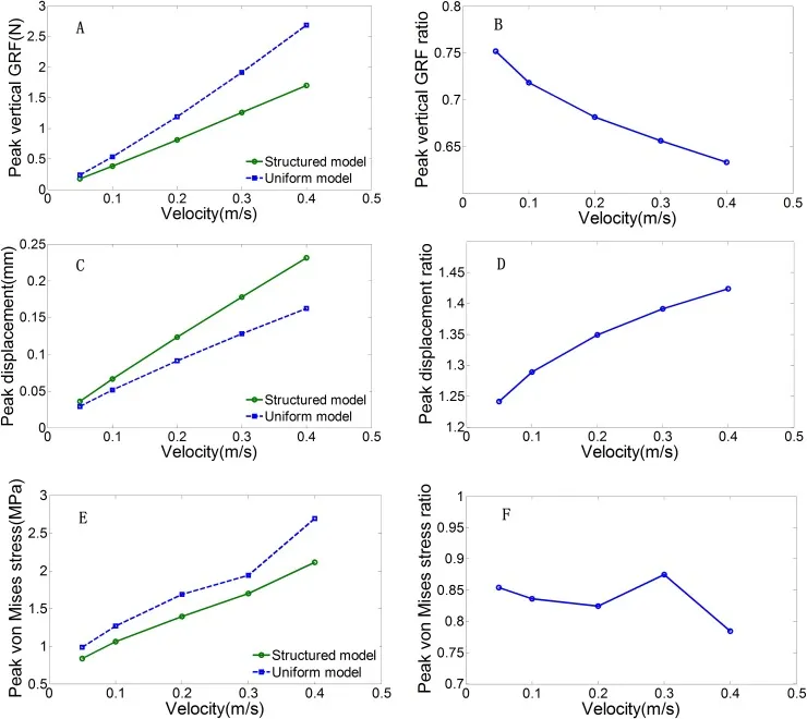 Peak GRF, displacement, and von Mises stress comparisons between structured and uniform models across impact velocities, with ratios showing structured superiority
Sensitivity tests varied dermal papillae stiffness (Young’s modulus 0.0004–4 MPa). Softer papillae enhanced GRF attenuation at high speeds but plateaued below 0.04 MPa.
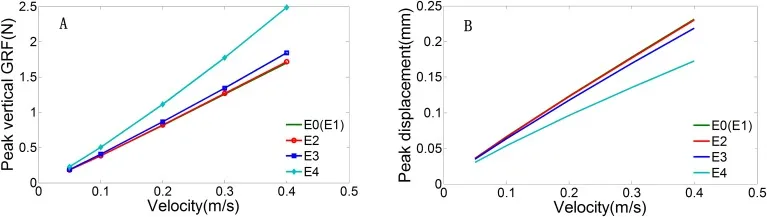 Peak GRF and displacement sensitivity to dermal papillae Young's modulus variations in dog paw pad FE models
For dogs in rugged activities, like biking, a reliable best dog bike trailer uk reduces unnecessary paw stress on hard surfaces.
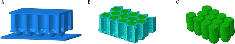 Micro-scale FE model assembly of dog paw pad epidermis, including stratified epithelium cube with dermal papillae cylinders, top plate, and ground
Material Properties Table:
| Component | Young’s Modulus (MPa) | Poisson’s Ratio | Element Type |
|---|---|---|---|
| Stratified Epithelium | 40 | 0.45 | C3D4 |
| Dermal Papillae | 0.004–4 | 0.49 | C3D4 |
| Top Plate | Rigid | – | C3D8R |
| Ground Plate | Rigid | – | C3D8R |
(Adapted from study data; friction coefficient 0.6 at pad-ground interface.)
Implications for Dog Health and Bio-Inspired Design
The epidermis offloads stress to tough epithelium, shielding soft inner layers—stresses drop ~1000-fold at the top. Dermis and subcutaneous act as energy absorbers, with fat compartments functioning like mini-hydrostatic cushions. All layers synergize for integrity during locomotion.
For pet care, this explains why breeds like German Shepherds excel in work: robust pads handle high GRFs. Owners should check pads regularly for cracks, foreign objects, or dryness—use vet-approved balms. Avoid extreme heat/cold; opt for best cooling blanket for dogs to prevent pad burns during summer runs.
This research inspires robotics (shock-absorbing feet) and human footwear, mimicking honeycomb for better impact protection.
In crates or travel, provide safe toys for puppies in crate to keep paws occupied without wear, and consider an x large heavy duty dog crate for larger breeds.
Conclusion
Dog paw pads’ micro-structure—honeycomb epidermis, collagen-rich dermis, and compartmentalized fat—delivers unmatched cushioning, reducing GRFs and stresses for safe, agile movement. Studies on German Shepherds and Chinese Rural dogs via SEM, histology, and FE prove this multi-layer synergy.
Protect your dog’s paws with routine checks, proper nutrition for skin health, and vet advice. Share your experiences in comments—have you noticed pad differences in breeds? Explore more on dog care for healthier pets.
References
- Miao H, et al. (2017). The micro-structure of digitigrade paw pads. Biology Open. DOI: 10.1242/bio.024828.
- Alexander RM, et al. (1986). Dynamic properties of mammalian footpads.
- Additional citations: Biewener (1990), Chi & Schmitt (2005), and NCBI PMC5769641 full bibliography.
Data: FigShare DOI:10.6084/m9.figshare.5620408. Approved by Jilin University IRB (20140418).