
Megaesophagus in dogs, a condition characterized by a dilated and poorly functioning esophagus, can be a profoundly distressing diagnosis for dog parents. The challenge of ensuring proper nutrition and preventing life-threatening complications often leaves owners feeling overwhelmed. However, advancements in veterinary medicine, including exciting New Treatment For Megaesophagus In Dogs, offer renewed hope. Integrative veterinarian Dr. Julie Buzby explains the causes, symptoms, diagnosis, and comprehensive management strategies for megaesophagus, empowering dog owners to provide the best possible care for their beloved companions.
Imagine a loyal, 7-year-old German Shepherd named Jasper, who, despite his usual patience, showed clear signs of distress. His dedicated mom described how he would immediately bring up all his food after eating, regardless of flavor or type. She worried constantly as Jasper lost weight and his energetic spirit diminished, wondering what his future held if he couldn’t even keep basic nourishment down. Based on these classic symptoms, Dr. Buzby suspected acquired megaesophagus, a condition that necessitates immediate attention and a well-informed approach to treatment. Understanding the intricacies of megaesophagus and exploring emerging therapeutic options is crucial for improving the quality of life for affected dogs.
 Chihuahua dog with head tilted, representing a pet owner's concern about canine health issues like megaesophagus
Understanding Canine Megaesophagus: What It Is and How It Affects Your Dog
Canine megaesophagus is a condition where the esophagus—the muscular tube connecting the mouth to the stomach—becomes abnormally enlarged or dilated and loses its ability to contract effectively. Normally, the esophagus actively pushes food and water down to the stomach through a series of coordinated muscle contractions called peristalsis. In a dog with megaesophagus, this vital process is severely compromised. Food gets trapped in the weakened, stretched-out esophagus instead of moving into the stomach for digestion. This obstruction leads to the primary and most noticeable symptom: regurgitation.
To illustrate this, picture a new pair of pantyhose—tight and efficient. Now, envision a stretched-out, flaccid leg of pantyhose. That’s similar to a megaesophagus: instead of being strong and narrow, the esophageal tube becomes weak and dilated, significantly hindering its function. This condition isn’t typically present from birth unless it’s congenital; rather, something disrupts the normal esophageal function, leading to its characteristic changes.
How a Dog’s Esophagus Should Work vs. What Happens in Megaesophagus
The journey of food from mouth to stomach is an active, complex process involving intricate muscle movements and nerve signals. When a dog swallows, food enters the upper esophagus. From there, a wave of muscle contractions (peristalsis) propels the food downwards, efficiently delivering it to the stomach. This coordinated action relies on healthy nerves sending precise messages to the esophageal muscles.
However, if these nerves fail to transmit the correct signals, or if there’s a physical obstruction (like a stricture or narrowing) preventing food passage, the esophagus begins to malfunction. Over time, the constant pressure of retained food and liquids causes the esophagus to dilate and lose its muscle tone, eventually resulting in megaesophagus. With impaired peristalsis, food cannot reach the stomach, leading to its passive expulsion, known as regurgitation. This often undigested food coming back up is usually the first red flag for concerned dog parents.
Regurgitation vs. Vomiting: Key Distinctions in Megaesophagus
Identifying regurgitation is critical for an early diagnosis of megaesophagus, as it differs significantly from vomiting. Regurgitation is a passive, effortless action. It occurs suddenly and without warning, often with no signs of nausea, gagging, or retching. The dog might simply lower their head, and food or water flows back out, appearing largely undigested because it never reached the stomach. This usually happens shortly after eating or drinking, though occasionally it can occur hours later. The material will not contain bile, which comes from the stomach and small intestine.
In contrast, vomiting is an active process. It involves visible effort, abdominal contractions, heaving, gagging, and retching. A dog might show signs of nausea (like excessive lip licking) before vomiting. The vomited contents can be partially digested, occur at varying times after eating, and may contain yellowish-green bile. Understanding these differences is paramount for accurately reporting symptoms to your veterinarian.
Types of Megaesophagus in Dogs: Congenital and Acquired Forms
Megaesophagus primarily presents in two forms: congenital and acquired. Each type has distinct characteristics, onset, and common causes.
Congenital Megaesophagus
Dogs born with megaesophagus have the congenital form. While it can affect any breed, it is more commonly observed in:
- German Shepherds
- Wire-Haired Fox Terriers
- Shar Peis
- Labrador Retrievers
- Miniature Schnauzers
- Newfoundlands
- Great Danes
- Irish Setters
Puppies typically begin showing signs of congenital megaesophagus around the time they transition from liquid mother’s milk to solid food, usually between weaning and 3-6 months of age. Milk, being liquid, can often flow down an abnormal esophagus more easily. In milder cases, symptoms may not become apparent until the dog is closer to one year old.
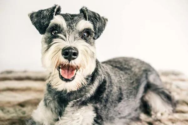 Miniature Schnauzer dog, a breed susceptible to congenital megaesophagus, looking bright and happy in an outdoor setting
Acquired Megaesophagus
Acquired megaesophagus develops later in a dog’s life, meaning the esophagus was normal at birth. Symptoms can emerge at any age, but it is most frequently diagnosed in middle-aged or senior dogs. This form is often a secondary symptom of an underlying disease or condition, making it crucial to identify the root cause for effective management.
Unraveling the Causes of Megaesophagus in Dogs
The etiology of megaesophagus is diverse, ranging from genetic predispositions in puppies to various systemic diseases in adult dogs.
Causes of Congenital Megaesophagus
- Incomplete Nerve Development: Some puppies are born with underdeveloped nerves responsible for esophageal function. Encouragingly, these nerves can sometimes mature as the puppy grows, leading to potential spontaneous resolution in 20-46% of cases.
- Vascular Ring Anomaly: This condition occurs when abnormal blood vessels, which usually disappear shortly after birth, persist and encircle the esophagus, constricting it. The most common form is a persistent right aortic arch (PRAA). This constriction prevents food from passing normally, causing the esophagus to dilate in front of the narrowed area.
Causes of Acquired Megaesophagus
- Myasthenia Gravis: This is the most common cause of acquired megaesophagus. It’s an autoimmune neuromuscular disease where the immune system attacks the receptors that transmit signals between nerves and muscles. As a result, the esophageal muscles don’t receive the “contract” message effectively, leading to dilation and dysfunction. Myasthenia gravis can also be a congenital condition.
- Geriatric Onset Laryngeal Paralysis and Polyneuropathy (GOLPP): Primarily affecting senior dogs (8-13 years), GOLPP is a degenerative neurological disorder. It typically involves three components: laryngeal paralysis (difficulty breathing due to impaired voice box function), megaesophagus, and hind limb weakness. The esophageal involvement stems from nerve degeneration.
- Trauma or Damage from a Foreign Body: Accidental ingestion of foreign objects (bones, toys, fish hooks) can cause direct trauma and inflammation to the esophageal tissue. If the body heals by forming scar tissue, it can create a stricture (narrowing) that obstructs food passage. Similar to a vascular ring anomaly, food accumulates above the stricture, causing the esophagus to dilate and lose function.
- Cancer: Tumors growing within or compressing the esophagus can lead to mechanical obstruction and subsequent dilation. Central nervous system cancers can also impair nerve function, contributing to megaesophagus.
- Miscellaneous Conditions: Several other conditions, though less common, can cause acquired megaesophagus:
- Myopathies: Diseases affecting the muscles themselves.
- Dysautonomia: Dysfunction of the autonomic nervous system.
- Central Nervous System Trauma: Injuries affecting the brain or spinal cord that impact nerve control of the esophagus.
- Botulism or Tetanus: Toxins that interfere with nerve-muscle communication.
- Addison’s Disease (Hypoadrenocorticism): An endocrine disorder affecting adrenal gland function.
- Bloat (GDV): Though primarily a stomach issue, severe bloat can put pressure on the esophagus.
- Esophagitis: Inflammation of the esophageal lining.
- Certain Toxins or Medications: Such as lead, organophosphates, or specific drugs like potassium bromide.
- Snake Bite: Can cause neurological or muscular damage.
- Hypothyroidism: While a link has been suggested, it’s not definitively proven as a direct cause.
Idiopathic Megaesophagus
In many cases, despite extensive diagnostic efforts, no underlying cause for megaesophagus can be identified. These are classified as idiopathic megaesophagus. This underscores the complexity of the condition and the need for comprehensive management strategies that address the symptoms regardless of the root cause.
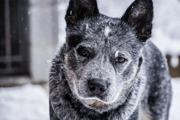 Senior Australian Cattle Dog (Heeler) enjoying the snow, illustrating a resilient dog despite potential health challenges like megaesophagus
Recognizing the Symptoms of Megaesophagus in Dogs
Early recognition of megaesophagus symptoms is vital for prompt veterinary intervention. Beyond regurgitation, several other signs can indicate the condition:
- Regurgitation: As discussed, this is the hallmark symptom—passive expulsion of undigested food or water, typically soon after eating.
- Weight Loss & Muscle Atrophy: Due to insufficient nutrient absorption, dogs with megaesophagus often lose significant weight despite a strong appetite. In chronic or severe cases, they may also exhibit noticeable loss of muscle mass.
- Difficulty Swallowing (Dysphagia): Dogs might struggle to get food down, displaying gulping, excessive head movements, or an abnormal neck posture during attempts to swallow, especially if an esophageal stricture or obstruction is present.
- Increased Drooling (Hypersalivation): While non-specific, some affected dogs may drool more than usual due to discomfort or inability to properly swallow saliva.
- Symptoms of Underlying Conditions: If megaesophagus is secondary to another disease, you might observe additional symptoms related to that condition. For example, a dog with GOLPP might have noisy breathing, while a dog with myasthenia gravis could show generalized muscle weakness that worsens with exertion.
- Symptoms of Aspiration Pneumonia: This is a severe and common complication where food, water, or saliva is accidentally inhaled into the lungs. Aspiration pneumonia is life-threatening and requires immediate veterinary care. Signs include:
- Wheezing or Labored Breathing: Harsh or difficult breathing sounds.
- Coughing and Gagging: A persistent, dry, or moist cough due to airway irritation.
- Rapid Breathing (Tachypnea) or Respiratory Distress: Fast breaths, stretching of the neck to breathe, or blue-tinged gums in severe cases.
- Fever: Elevated body temperature (above 102.5°F or 39.2°C).
- Nasal Discharge: Thick, yellowish-green snot.
- Lethargy: Reduced energy, sluggishness, and reluctance to engage in normal activities.
If you suspect regurgitation, unexplained weight loss, or any of the above symptoms (especially those indicating aspiration pneumonia), contact your veterinarian promptly. Aspiration pneumonia constitutes a veterinary emergency.
Diagnosing Megaesophagus and Its Underlying Causes
A thorough diagnosis begins with a detailed history of your dog’s symptoms and a comprehensive physical examination. If megaesophagus or secondary aspiration pneumonia is suspected, the initial diagnostic steps typically involve:
Chest X-rays (Radiographs): This is often the quickest and easiest way to identify esophageal dilation and check for aspiration pneumonia. In some cases, a liquid contrast agent (administered with extreme caution due to aspiration risk) can be given orally to highlight the esophagus and reveal subtle abnormalities.
Fluoroscopy: This real-time X-ray procedure allows a veterinary specialist to observe the movement of food and liquids down the esophagus, providing invaluable information on esophageal motility issues that may not be evident on static X-rays.
Identifying the Root Cause
Once megaesophagus is confirmed, the next crucial step is to pinpoint the underlying cause, particularly for acquired forms. Diagnostic tests vary depending on the suspected condition:
- Blood Tests: To screen for Addison’s disease, hypothyroidism, and signs of infection or inflammation (e.g., from aspiration pneumonia).
- Sedated Laryngeal Examination: To assess for laryngeal paralysis, a key component of GOLPP.
- Specific Blood Tests or Tensilon Test: To confirm myasthenia gravis by evaluating the immune system’s attack on nerve-muscle junctions or the response to a specific medication.
- Endoscopy: Involves inserting a flexible tube with a camera into the esophagus to visually inspect for strictures, foreign bodies, tumors, or esophagitis.
- Advanced Imaging (CT/MRI), Biopsies, Nerve Stimulation Tests: May be recommended by specialists if initial tests are inconclusive.
Despite these advanced diagnostic tools, some cases remain idiopathic, meaning the underlying cause cannot be determined. Even in such instances, effective management strategies can significantly improve a dog’s quality of life.
Navigating Treatment for Megaesophagus: Embracing New Hope
While finding and treating the underlying cause is ideal, many dogs with megaesophagus can be effectively managed symptomatically, regardless of whether a specific cause is identified. Treatment focuses on two main pillars: minimizing regurgitation and addressing the primary disease (if found).
Strategies to Minimize Regurgitation
The cornerstone of megaesophagus management lies in specialized feeding techniques and, increasingly, novel pharmacological approaches.
Optimizing Food Consistency and Frequency:
Different dogs respond best to different food consistencies. Your veterinarian will help determine if your dog fares better with liquids, slurries, or solids.- For dogs struggling with solids: Liquid diets or slurries (food blended with water or broth) are often recommended. Higher water content foods can also be beneficial.
- For dogs struggling with liquids: Solid food, often formed into small “meatballs” from canned food, can be more effective. The right consistency and size can sometimes stimulate enough esophageal activity to aid passage into the stomach.
- Small, frequent meals: This strategy reduces the volume of food in the esophagus at any one time, lessening the likelihood of regurgitation.
- High-calorie foods: These provide adequate nutrition with smaller portions, further easing the esophageal burden.
Elevated Feeding with a Bailey Chair:
Gravity is your ally in managing megaesophagus. Elevated feeding positions help food move downwards into the stomach.- Basic Elevation: Placing food and water bowls on a raised platform (stool, staircase) to encourage your dog to eat while standing on their hind legs.
- The Bailey Chair: This specialized feeding chair, resembling a high chair for dogs, is highly recommended. It keeps the dog in an upright, vertical position during and after meals. This position maximizes gravitational assistance and is crucial for preventing regurgitation. Dogs should remain in the Bailey Chair, or confined and upright, for at least 10-15 minutes after eating or drinking to allow gravity to do its work.
 Senior Boxer dog being fed in an elevated position by an owner, demonstrating a key management strategy for dogs with megaesophagus
Feeding Tubes:
In severe cases where oral feeding strategies are insufficient or aspiration pneumonia is recurrent, a feeding tube (e.g., a gastrostomy tube) may be surgically placed. This allows food to bypass the esophagus entirely and deliver nutrients directly to the stomach. While highly effective for nutrition, it’s important to remember that feeding tubes do not prevent regurgitation of saliva, which dogs naturally swallow throughout the day.Administering Sildenafil: A Promising New Treatment for Megaesophagus in Dogs
One of the most exciting recent developments in megaesophagus management is the potential use of the human drug sildenafil (commonly known as Viagra®). Historically used for pulmonary hypertension, sildenafil is a phosphodiesterase-5 (PDE5) inhibitor. Its mechanism of action involves relaxing smooth muscles, and research has shown it can relax the lower esophageal sphincter (LES) in dogs. The LES is a ring of muscle at the bottom of the esophagus that normally relaxes to allow food into the stomach and tightens to prevent stomach contents from refluxing back up. In megaesophagus, impaired LES relaxation can exacerbate food retention.A groundbreaking 2022 study published in the American Journal of Veterinary Research evaluated the effects of compounded liquid sildenafil compared to a placebo in 10 dogs with megaesophagus. The results were promising:
- Reduced Regurgitation: While individual responses varied, dogs on sildenafil showed a significant reduction in the frequency of regurgitation compared to when they received a placebo.
- Weight Gain: Consistent with reduced regurgitation, dogs also gained more weight while on sildenafil, indicating improved nutrient absorption.
- Mechanism: Researchers hypothesize that sildenafil’s ability to relax the lower esophageal sphincter allows food to pass more easily from the esophagus into the stomach, mitigating the pooling effect.
Despite the small sample size and challenges noted by researchers in administering sildenafil to dogs with poor esophageal motility, this study represents a significant step forward. Sildenafil offers a novel pharmacological approach that directly addresses a physiological component of megaesophagus. While more research is needed to fully understand its long-term efficacy and ideal dosing protocols, it provides a valuable new treatment for megaesophagus in dogs and a beacon of hope for improving patient outcomes. Veterinarians are increasingly exploring its use as an adjunctive therapy alongside traditional feeding strategies.
 Senior Boxer dog being fed in an elevated position by an owner, demonstrating a key management strategy for dogs with megaesophagus
Addressing the Underlying Cause
If an underlying cause for acquired megaesophagus is identified, specific treatments are initiated:
- Myasthenia Gravis: Treated with immunosuppressants (like corticosteroids) and anticholinesterase inhibitors (e.g., pyridostigmine) to improve nerve-muscle transmission.
- Vascular Ring Anomaly: Surgical correction is typically performed in puppies to remove the constricting vessel. While surgery can improve symptoms, complete resolution of megaesophagus occurs in only about 30% of cases.
- Esophageal Stricture: May be treated with balloon dilation (endoscopically guided stretching) or stent placement.
- Infections/Inflammation: Appropriate antibiotics or anti-inflammatory medications.
- Endocrine Disorders: Management of conditions like Addison’s disease or hypothyroidism.
Prognosis for a Dog with Megaesophagus
The prognosis for megaesophagus in dogs generally remains guarded, and it is often a lifelong condition. The main exception is some puppies with congenital megaesophagus (not caused by a vascular ring anomaly), whose esophageal function may spontaneously improve by around six months of age.
For acquired megaesophagus, the outcome heavily depends on identifying and successfully treating the underlying cause. While some underlying conditions like myasthenia gravis can lead to significant improvement or even resolution of megaesophagus with proper treatment, there are no guarantees. Unfortunately, current medications or surgical options, even for vascular ring anomalies, do not always fully restore normal esophageal function.
The most significant factor contributing to the poor prognosis is the persistent risk of aspiration pneumonia. This life-threatening complication can cause severe lung damage and recurrence, greatly impacting a dog’s quality of life and longevity. Even with diligent care, dogs with megaesophagus require constant vigilance for signs of respiratory distress.
However, the emergence of new treatment for megaesophagus in dogs, like sildenafil, offers a glimmer of hope. While not a cure, such medications, combined with meticulous feeding management and a strong partnership with your veterinarian, can significantly improve a dog’s comfort, reduce regurgitation, and enhance overall quality of life.
Jasper’s Journey: Living with Megaesophagus
For Jasper, the German Shepherd patient, the specific cause of his acquired megaesophagus remained undetermined. His dedicated mom, however, didn’t let that deter her. She custom-built a Bailey Chair, which Jasper now uses for every meal, eager for his food. Through consistent elevated feeding and carefully monitored mealtimes, Jasper gradually regained the weight he had lost. His mom diligently watches for any signs of aspiration pneumonia, knowing that vigilance is key to managing his condition. Her quick communication with Dr. Buzby ensures that any concerns are addressed promptly.
Managing Megaesophagus: A Partnership for Hope and Health
A diagnosis of megaesophagus is undoubtedly concerning, and the journey can be challenging. However, dog parents play a pivotal role in managing this condition. Much of the successful care revolves around implementing consistent, appropriate feeding strategies tailored to your dog’s needs. Adhering strictly to your veterinarian’s feeding plan can dramatically reduce the risk of regurgitation and the dreaded complication of aspiration pneumonia.
While the specialized feeding routine, especially with a Bailey Chair, might initially feel cumbersome, it also creates an invaluable opportunity for quality bonding time with your dog. Those 10-15 minutes of upright post-meal stillness can be filled with gentle petting, reassuring words, and ear scratches, transforming a medical necessity into a loving ritual.
Establishing a robust partnership with your veterinarian is also non-negotiable. An experienced vet who is intimately familiar with your dog’s megaesophagus case can make timely adjustments to the treatment plan and provide swift intervention if aspiration pneumonia arises. Together, with dedication, informed choices, and the potential benefits of new treatments like sildenafil, you and your vet can work to ensure your dog experiences the happiest and most comfortable life possible despite megaesophagus.
References
- American Journal of Veterinary Research, 2022. “Effects of compounded liquid sildenafil vs a placebo in 10 dogs with megaesophagus.”
Note: This article is for informational purposes only and does not constitute medical advice. Always consult with a qualified veterinarian for any health concerns or treatment decisions regarding your pet.