
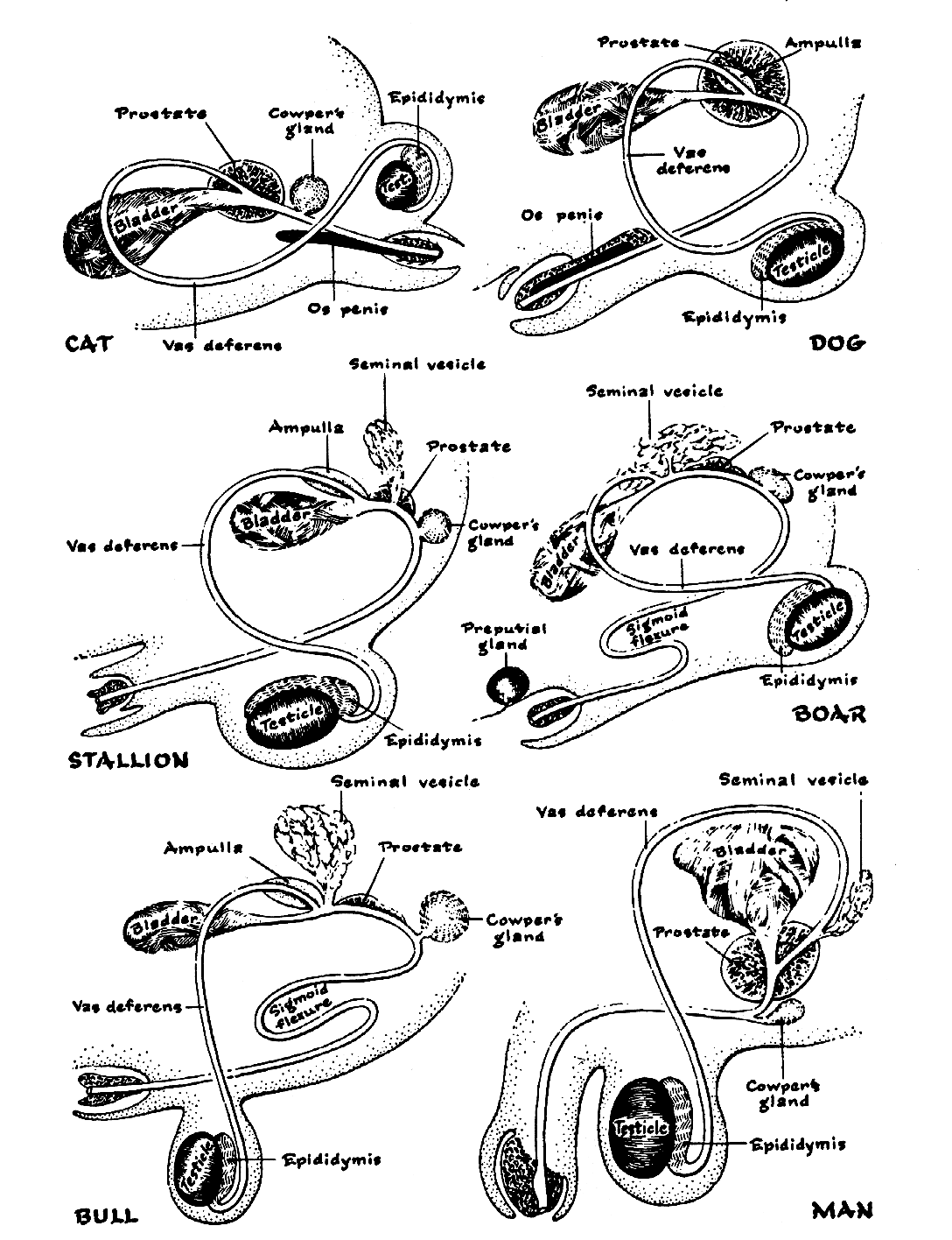
The male reproductive systems of dogs and cats, while sharing fundamental similarities, exhibit distinct anatomical differences that are crucial for understanding their reproductive functions. These systems are intricately designed to produce spermatozoa through spermatogenesis, secrete fluids that support sperm viability and transport, and produce testosterone, the hormone essential for developing male characteristics and behaviors. This article delves into the anatomy and function of these systems, highlighting key distinctions between the canine and feline species.
Anatomy and Function of the Male Reproductive System
The primary functions of the male reproductive system in both dogs and cats are:
- Spermatogenesis: The continuous production of sperm, which are capable of fertilizing the female ovum.
- Fluid Secretion: The production of seminal fluids that aid in sperm survival and facilitate their transport into the female reproductive tract during coitus.
- Hormone Production: The secretion of testosterone, a hormone vital for the development of secondary sexual characteristics and influencing male behavior patterns.
The Testis and Scrotum
A pair of testes is responsible for sperm production. Spermatogenesis optimally occurs at temperatures below 40°C. In adult animals, the testes are housed externally within the scrotum, a pouch of pigmented, sparsely furred skin. In dogs, the scrotum is located between the upper thighs, while in cats, it is positioned ventrally to the anus, near the ischial arch.
The scrotum’s temperature regulation is managed by the Dartos muscle. In cold conditions, this muscle contracts, drawing the testes closer to the body for warmth. Conversely, in warmer weather, it relaxes, allowing the scrotum and testes to descend, thereby facilitating cooling.
Internal Structure of the Testis
The testicular tissue is primarily composed of coiled seminiferous tubules. These tubules are lined by spermatogenic cells, which are responsible for sperm formation, and Sertoli cells, which provide nutrients to prolong sperm life and secrete small amounts of estrogen. Interspersed between the seminiferous tubules are the Leydig cells, also known as interstitial cells, which are the primary source of testosterone production.
The seminiferous tubules converge to form the epididymis, a long, coiled tube situated along the dorso-lateral border of the testis. The epididymis, particularly the cauda epididymis at the caudal extremity of the testis, serves as the site for final sperm maturation and storage before ejaculation.
Spermatic Cord and Tunica Vaginalis
The epididymis continues as the deferent duct (vas deferens or ductus deferens), which exits the scrotum and enters the peritoneal cavity via the inguinal ring. Within the scrotum, the testis is enveloped by the tunica vaginalis, a double-layered serous membrane derived from the peritoneum. This membrane also encloses the spermatic cord, which contains the testicular artery, vein, nerves, and the deferent duct. The testicular artery forms the pampiniform plexus, a network of arterioles that cools the blood before it reaches the testicular tissue, ensuring optimal conditions for spermatogenesis. The cremaster muscle, located within the spermatic cord, works in conjunction with the Dartos muscle to adjust scrotal position and temperature.
Deferent Duct and Urethra
The deferent duct, a continuation of the cauda epididymis, travels within the spermatic cord and enters the peritoneal cavity through the inguinal ring. In both dogs and cats, the deferent ducts join the urethra within the prostate gland.
The urethra serves as a common passageway for both the urinary and reproductive systems, extending from the bladder’s neck to its external opening at the tip of the penis.
Urethral Differences in Dogs and Cats
Significant differences exist in the urethral structure between dogs and cats:
- Dog: The canine urethra is lengthy, comprising a pelvic part along the floor of the pelvis and a penile part running through the penis, with its opening directed cranially.
- Tomcat: The tomcat possesses a short preprostatic urethra between the bladder neck and the prostate gland. The penile urethra is considerably shorter, extending only to the ischial arch, with its opening directed caudally and located ventral to the anus. This caudal orientation is associated with the tomcat’s territorial marking behavior, where they spray urine onto vertical surfaces.
Accessory Glands
Both species possess a prostate gland located near the bladder neck. Cats also have bulbo-urethral glands situated near the tip of the penis. These glands contribute secretions that increase ejaculate volume, aiding in sperm propulsion. Their alkaline pH neutralizes any residual urine in the urethra, creating a more favorable environment for sperm survival.
The Penis
As the urethra exits the pelvic cavity, it becomes encased by erectile tissue, forming the corpus cavernosum penis. This tissue, composed of endothelium-lined spaces, engorges with blood under pressure during sexual excitement, leading to erection and enabling intromission into the female vagina.
Penile Differences in Dogs and Cats
- Dog: The canine penis is anchored to the ischial arch by muscular crura, which merge to form the penile root. It then curves cranioventrally, forming the body and the glans penis. A small bone, the os penis, is embedded within the glans penis dorsal to the urethra. This bone aids in maintaining rigidity during the initial stages of coitus before full erection and vaginal clamping by the bitch. The urethra passes through a bony tunnel within the os penis, which can be a site for urethral calculi blockage due to its inability to expand.
- Tomcat: The tomcat’s penis features a shorter segment of erectile tissue where the bulbo-urethral glands open. Ventral to the urethra within the erectile tissue lies a bony os penis. The urethra is narrowest in this region, making it prone to blockage by struvite crystals. The glans penis is adorned with small, backward-pointing barbs. Upon withdrawal after coitus, these barbs cause intense pain to the queen, often eliciting a loud howl. This pain triggers a reflex arc that, approximately 36 hours later, results in ovulation, classifying the queen as an induced ovulator.
Prepuce
The penis of both dogs and cats retracts into a protective sheath called the prepuce when relaxed. The outer surface is covered in hairy skin, while the inner lining is a mucous membrane continuous with the urethra, richly supplied with lubricating glands. Preputial infections, known as balanoprosthitis, can lead to an unpleasant greenish discharge.
Author: Victoria Aspinall bvsc mrcvs
To cite this article use either:
DOI: 10.1111/j.2045-0648.2010.00025.X or Veterinary Nursing Journal Vol 26 pp89-91
Bibliography:
ASPINALL, V. and CAPELLO, M, 2009 Introduction to Veterinary Anatomy and Physiology 2nd ed. Butterworth Heinemann. Oxford.
DYCE, K. M., SACK, W. O. and WENSING. C. J. G. 2002 Textbook of Veterinary Anatomy 3rd ed Saunders. Philadelphia.
EVANS. H. E. 1993 Miller’s Anatomy of the Dog 3rd ed. Saunders. Philadelphia.
Veterinary Nursing Journal • VOL 26 • March 2011 •